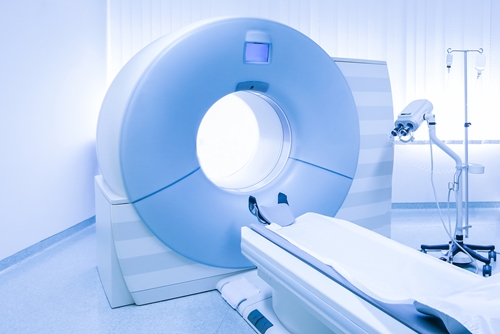
On Nov. 6 researchers from Massachusetts General Hospital published a medical-imaging study. The research claims magnetic resonance imaging can effectively pre-select stroke patients for catheter-based blood clot treatments, reported EurekAlert.
During the study, which was published in the journal JAMA Neurology, doctors evaluated the ischemic core – the part of the brain most often damaged during ischaemic attacks, or mini-strokes – in 103 patients to determine if they would benefit from a blood clot-busting procedure. Physicians used a combination of computed tomographic angiograph and MRI scans to assess 72 of the test subjects. The remaining 31 underwent only CT scans.
The 72 participants who received dual scans were divided into two post-scan categories: likely to benefit and uncertain to benefit. Ultimately, 40 were identified as LTB and 32 were labeled UTB.
After a three-month, post-treatment recovery period, physicians revaluated both groups. In the LTB group, 52 percent of the patients regained full function. Additionally, 32 percent of stroke patients classified UTB fully recovered. In the group that received only CT scans, 31 percent of participants were also able to make a full recovery.
Though this data was initially encouraging, the MGH researchers uncovered even more exciting results when they compared their work to other stroke-related medical-imaging research. In two earlier studies, scientists found that 7 to 13 percent of patients who underwent only post-stroke CT scans received endovascular treatments. In the MGH study, 33 percent of patients who received both CT scans and MRIs were cleared for treatment. This suggested that MRIs provided a clearer image than CT scans and, in turn, allowed doctors to select more patients for treatment.
"The recent ground-breaking endovascular clinical trials used CT selection criteria for stroke patients as an effective way to identify who would benefit from endovascular stroke therapy," the study's lead author Dr. Thabele Leslie-Mazwi told EurekAlert. "Our work shows that MRI identifies patients with greater precision, which means that fewer patients are inaccurately excluded from a treatment that might otherwise benefit them."
Scanning For Stroke Patients
Strokes occur when cerebral blood flow is interrupted by blood vessel blockages – usually via clots – or ruptures. This causes a variety of symptoms, including confusion, facial paralysis and vision loss.
According to the American Stroke Association, stroke victims must undergo screenings – usually within three to six hours after the event – so that doctors can assess the brain damage caused by the stroke. Physicians also use these scans to determine if victims should receive endovascular treatments or a tissue plasminogen activator, a drug that can re-open blocked cerebral blood vessels. Both of these treatments are potentially deadly, as they can cause brain hemorrhages in stroke victims with ruptured cerebral blood vessels.
CT Advancement
In recent years, researchers have focused on evaluating the CT scan and its place in the realm of stroke prevention and treatment. In 2008 a group of Spanish doctors presented a study at Radiological Society of North America's annual meeting that claimed the CT scan was the best scanning tool for emergency physicians looking to screen patients for post-stroke treatment. The research was later published in the journal RadioGraphics.
Last year Canadian researchers also found that CT scanning greatly increased a physician's ability to treat individuals who suffered from ischaemic attacks, reported the British Broadcasting Corporation.
MRI Makes Headway
In 2006 researchers at Stanford University found that MRIs could give doctors a nuanced view of the post-stroke brain, the university said in a news release. Dr. Greg Albers and his team studied the pre- and post-treatment MRI scans from 74 stroke patients. Three patients died from fatal brain hemorrhages after treatment. Using the scans from these deceased patients, Albers was able to identify a high-risk MRI archetype. He also found that 67 percent patients who were identified as likely to benefit from treatment ultimately recovered.
"By having this additional information available, we should be able to make a much more sophisticated decision about which therapies are optimal for an individual patient, especially as you get into the longer time windows," Albers told Stanford University.
Over the years, numerous other studies have shown the benefits that accompany post-ischaemic stroke MRI scans. Preventive organizations have responded accordingly. In 2010, the American Academy of Neurology amended its stroke scanning guidelines to reflect this trend.
"While CT scans are currently the standard test used to diagnose stroke, the Academy's guideline found that MRI scans are better at detecting ischemic stroke damage compared to CT scans," Dr. Peter Schellinger, lead author of the AAN's guidelines, said in a press release.
Ronny Bachrach
Latest posts by Ronny Bachrach (see all)
- Konica Minolta Debuts First-of-Its-Kind Digital U-Arm System at AHRA - July 27, 2016
- Researchers Detect Signs Of Stroke Risk Using MRI - June 27, 2016
- Imaging Biz: Q&A with David S. Channin MD: How to Make PACS Patient Centered - June 22, 2016


