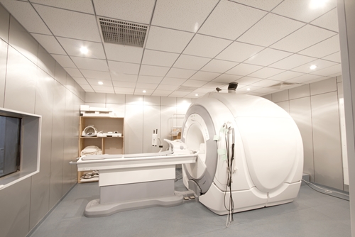
Canadian researchers may have uncovered a new technique for detecting early signs of stroke risk in diabetics. A team of medical-imaging analysts at Sunnybrook Research Institute in Toronto used 3-D magnetic resonance imaging to find plaque in the carotid arteries of individuals with Type 2 diabetes, reported AuntMinnie.
According to Johns Hopkins Medicine, physicians normally assess stroke risk by using a conventional MRI or other clinical tests to look for narrowing in the carotid arteries, the vessels on the neck that pump blood to the brain. Strokes can occur when these arteries narrow too much and collapse. However, this risk-assessment technique does not work for individuals who fail to display clear indications of carotid artery stenosis.
For the study, presented at the 2015 annual meeting of the Radiological Society of North America, researchers scanned 159 asymptomatic diabetes patients for traces of intraplaque hemorrhaging or a slight build up of plaque in the carotid arteries. None of them displayed signs of stenosis. The average age of the test group was 63.
Ultimately, 37 of the subjects showed signs of IPH. Five had IPH in both carotid arteries. Additionally, all subjects also displayed signs of artery wall thickening.
"We are hoping that the presence of IPH in 23% of our population warrants a clinical trial or even a change in clinical guidelines to take IPH into account when treating patients," the study's lead author Dr. Tishan Maraj said in an interview with the news source. "We are hoping that this study or future studies can influence that."
Traditional Risk Detection
Most doctors look for blockages by listening to carotid arteries with a stethoscope, assessing arterial blood flow via duplex ultrasound scans or collecting images using MRI or magnetic resonance angiography. However, these tests only effectively gauge stroke risk for patients who exhibit symptoms of carotid artery stenosis. Last year the U.S. Preventive Services Task Force even advised medical professionals to discontinue testing asymptomatic individuals for CAS. Overdiagnosis and screening ineffectiveness were the primary drivers behind the USPSTF ruling. The organization published a number of studies to support its claims. For instance, in 2007, USPSTF researchers put out a study that argued ultrasound was an ineffective testing measure for asymptomatic individuals.
This trend has lessened the screening options for at-risk adults without explicit signs of CAS. Additionally, preventive testing continues to gain importance as rates of mortality due to cardiovascular or circulatory disease rise. According to The New England Journal of Medicine, the number of deaths due to these diseases rose by 41 percent from 1990 to 2013.
The Advantages Of 3-D Imaging
Maraj and his team used 3-D MRI to spot thin layers of buildup sticking to carotid walls. According Maraj, this scanning method allows analysts to capture a three-dimensional image of the entire artery in one session. To achieve a similar result with a traditional MRI, imaging professionals must collect a series of two-dimensional scans and piece them together to form a complete picture of the artery.
Contact Viztek for more information.
Ronny Bachrach
Latest posts by Ronny Bachrach (see all)
- Konica Minolta Debuts First-of-Its-Kind Digital U-Arm System at AHRA - July 27, 2016
- Researchers Detect Signs Of Stroke Risk Using MRI - June 27, 2016
- Imaging Biz: Q&A with David S. Channin MD: How to Make PACS Patient Centered - June 22, 2016


