Recent debates have been heating up in the medical imaging industry regarding the effectiveness of various breast screening modalities. In the place of traditional mammography, physicians are beginning to utilize the innovative method of digital breast tomosynthesis to generate highly detailed scans of breasts. But what is the true effectiveness of Digital Breast Tomosynthesis ?
Similar to low-dose Mammography Equipment, Tomosynthesis uses decreased dosages of X-rays to develop a three-dimensional view of the breast. This provides radiologists and other imaging specialists with a more comprehensive idea of breast density, which can be considered a risk factor for breast cancer development in women. The conventional, two-dimensional mammography scans may mask suspicious or potentially malicious growths that are missed due to overlapping tissue in the scans. Much like digital radiography, tomosynthesis allows radiologists to see faster results and thusly provide better treatment plans to patients.
Testing DBT
At last year’s Radiological Society of North America convention, a team of researchers from Massachusetts General Hospital presented results from a significant tomosynthesis study. Led by Pragya Dang, M.D., the scientists reviewed more than 170 biopsy-proven breast cancer tumors that were collected between March 2011 and October 2012.
Using tomosynthesis and conventional mammography, the radiologists imaged the cancerous tumors and determined the efficacy of both modalities. Out of all the samples, only 3 percent were hidden from the view of tomosynthesis. Compared to 16 percent missed through digital mammography, the results showed how much more thorough and beneficial tomosynthesis may be than the traditional methods.
“There was a significantly improved degree of visibility with tomosynthesis compared to digital mammography,” Dang said during her presentation.
Working with DBT
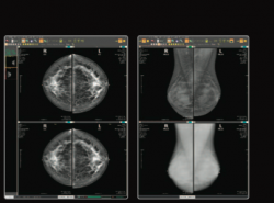
Sometimes referred to as 3D mammography, tomosynthesis digital imaging partially rotates around the breast and captures images from different angles. Typically, the scanner will take between 10 and 15 images per procedure. Afterwards, the images are uploaded into a computer program, such as a PACS, that assembles the scans into one comprehensive 3D image of the breast.
Studies, such as the one conducted by Dang’s team at Massachusetts General, have shown that tomosynthesis can improve the visualization of tumors, reduce recall rates and improve the identification of tissue. As such, 3D mammography can help identify malignant lumps in breasts well before they can be physically felt by a patient. Early detection is the key to increasing survival rates for women who may be diagnosed with breast cancer, as they might have more treatment options available to them.

Latest posts by Steve Deaton (see all)
- Practice Better Radiology to Avoid Malpractice Lawsuits - July 16, 2014
- Medical Imaging Data Can Help Improve the Patient Experience - June 19, 2014
- Replace Outdated PACS Systems to Experience Cost Savings in Imaging - June 16, 2014
 0
0 0
0 0
0

