The mere mention of cancer creates fear and anxiety in many patients. Medical imaging can show where potentially cancerous cells are, but blood tests and biopsies are needed to determine whether they are benign or malignant. This much testing does nothing to calm patients' uneasiness over the situation. However, a new study explained a way to diagnose cells with less invasive procedures.
Researchers use MRIs for clarification
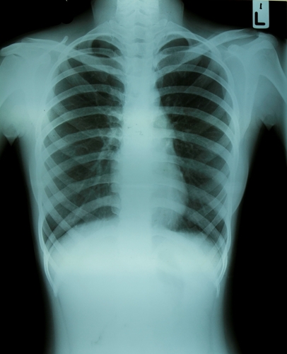
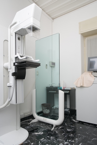
In traditional cancer diagnoses, digital imaging has been used to get a glimpse of the growth and see if any disease has spread. However, a new study published in the journal Nature Communications shows MRIs can be used for much more. Researchers at John Hopkins University discovered a method that uses the device to determine whether cells are cancerous. Without needing to draw blood or take tissue samples of the infected area, doctors will be able to diagnose patients with the disease.
Healthy cells have sugar attached to mucin, which shows up as a deep red in MRIs. However, cancerous cells have fewer of these sugary proteins, so that crimson color is missing, AuntMinnie.com explained. This method allows radiologists to utilize cells' properties instead of injecting patients with contrast dyes. While this testing has only been explored in test tubes and mice, it shows promise in humans.
New method improves safety and convenience
Current examinations of potentially cancerous cells require invasive procedures and extensive imaging to diagnose the disease. As stated in the source, the researchers' new use for MRI machines could be used to detect early-stage cancer and possibly eliminate biopsies altogether, which would be convenient for patients and doctors.
In addition to producing a more complete image, this method also prevents potentially harmful chemicals from entering patients' bodies. Researchers from the Mayo Clinic found that gadolinium contrast dyes remain in the brain years after MRI testing, as reported in their study in the journal Radiology. These chemicals have also been linked to nephrogenic systemic fibrosis in dialysis patients, AuntMinnie.com explained. If approved for human use, John Hopkins' MRI scanning will eradicate the use of gadolinium-based contrast agents in diagnostic imaging for cancer.
Using the body's already existing characteristics to determine malignancy and harmlessness in cells provides an inexpensive and efficient way to diagnose cancer in patients. By not exposing people to GBCAs, physicians can use MRI scans on all patients, even those who already have the disease, without causing more harm.
Contact Viztek for more information.

Ronny Bachrach
Latest posts by Ronny Bachrach (see all)
- Konica Minolta Debuts First-of-Its-Kind Digital U-Arm System at AHRA - July 27, 2016
- Researchers Detect Signs Of Stroke Risk Using MRI - June 27, 2016
- Imaging Biz: Q&A with David S. Channin MD: How to Make PACS Patient Centered - June 22, 2016
 0
0 0
0 0
0