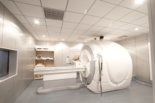
Medical imaging in the emergency department has a proven track record of enhancing diagnoses and treatment strategies. Now, oncologists at the University of San Diego Moores Cancer Center are using MRI in combination with traditional ultrasound prostate exams to provide more precise cancer care.
FierceMedicalImaging reported that the physicians, producing three-dimensional images of the prostate, were able to identify previously undetectable growths. The team was led by J. Kellogg Parsons, M.D., a urologic oncologist who helped develop the new procedure. The technique has patients undergo an MRI before participating in a biopsy exam. The images are combined with real-time results from the ultrasound to create a "road map" of the prostate.
"With an ultrasound exam, we are typically unable to see the most suspicious areas of the prostate, so we end up sampling different parts of the prostate that statistically speaking are more likely to have cancer," said Parsons, quoted by the news source. "The MRI is a game-changer. It allows us to target the biopsy needles exactly where we think the cancer is located. It's more precise."
With the biopsy guided by MRI, the patient experience is enhanced by reducing instances of false positives. Doctors can provide earlier diagnosis when cancer is present, which can get patients started on treatment plans as soon as possible.
Use of MRI, CT down in EDs
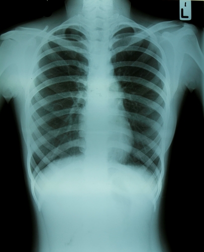
While Kellogg and his colleagues demonstrated the effectiveness of MRI, a new study showed that the use of diagnostic imaging in emergency departments has dropped since 2007. Published in the American Journal of Roentgenology, researchers from the Brigham and Women's Hospital examined the use of scans from January 1993 through December 2012.
Led by Ali Raja, M.D., the team found that the number of ED visits during that time span increased 28 percent, from roughly 48,000 to 61,000 per year. They also monitored the severity of illness for ED visits and categorized the number of imaging studies based on modality – X-ray, ultrasound, MRI, CT and others. Researchers also measured each exam's associated relative value units (RVUs).
During the nearly 20-year time frame, Raja and his colleagues found that total imaging RVUs increased by 267 percent from 1993 to 2007. However, by 2012, the rate had reduced by 19.2 percent. The actual number of digital imaging procedures during the early time period rose 66.5 percent, but dropped 17 percent after 2012.
Out of all the measured modalities, CT and MRI experienced some of the largest decreases in utilization after 2007 at 33.4 percent and 20.6 percent, respectively. The drop in use can be attributed to growing concerns over protecting patients from radiation exposure, but it might not be the sole cause. The two procedures have relatively high costs in comparison to other modalities, which could lead to fewer procedures, but increased focus on determining the appropriateness of certain exams may be the main reasoning behind the lower utilization numbers. As a result, decision-support tools have grown in popularity to guide radiologists on ordering exams.
Contact Viztek for more information.

Ronny Bachrach
Latest posts by Ronny Bachrach (see all)
- Konica Minolta Debuts First-of-Its-Kind Digital U-Arm System at AHRA - July 27, 2016
- Researchers Detect Signs Of Stroke Risk Using MRI - June 27, 2016
- Imaging Biz: Q&A with David S. Channin MD: How to Make PACS Patient Centered - June 22, 2016
 0
0 0
0 0
0