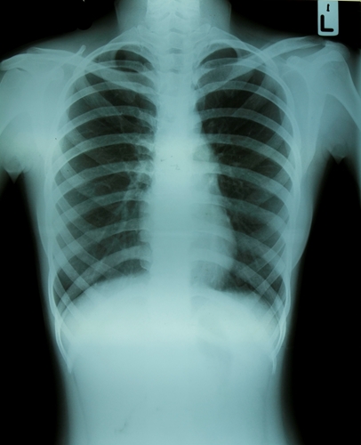
After sustaining an injury to the wrist, many patients wind up undergoing medical imaging exams to determine severity. The sooner that radiologists can identify symptoms of sprains, breaks or fractures, the quicker they can set individuals up with recovery plans.
According to Heathline, wrist fractures occur when the radius bone in the forearm cracks or breaks. It is also known as a transverse wrist fracture or distal radius fracture. Depending on how severe the fracture is, patients can experience varying levels of pain. Many people sustain this injury when putting a hand out to stop themselves from falling, but older patients are more susceptible due to their bones' fragility.
At times, wrist fractures can be set and wrapped in a cast until the bone has healed. However, serious breaks might require surgery to properly repair the bones.
Recently, researchers from Dokuz Eylul University in Izmir, Turkey, conducted a study on the use of radiography in scanning for wrist fractures. The results were published online in the October issue of Emergency Radiology. Led by Ali Balci, Ph.D, the scientists believed that physicians' inclinations toward CT and MRI are causing them to miss approximately one-third of wrist fractures.
According to AuntMinnie.com, traditional diagnostic radiology has been the norm for identifying injuries in the emergency room, but multiple detector CT has demonstrated high-quality resolution on images that depict fractures with ease. Balci and his team wanted to test the modalities' efficacy with a large patient population to yield optimal results.
Narrowing down the benefits of MDCT
The Dokuz researchers started by reviewing patients in the ER who underwent both radiography and CT exams for wrist injuries. They found that 455 patients, which included 264 men and 191 women, met the study's criteria.
Using a 16-detector-row CT scanner, the researchers had patients receive MDCT within 72 hours of trauma. Two experienced radiologists performed readings without knowing the modality's findings, searching for presence of fractures. Forty-nine percent of the patient population had one or more fractures, with a total of 457 wrists being broken. MDCT found 51 percent of negative fractures in 232 patients.
On the other hand, traditional radiography yielded 57.8 percent sensitivity on all wrist fractures and 38.7 percent for carpal fractures, which occur in the proximal pole or distal tubercle, Medscape explained. In addition, 47 of the scans were considered to be suboptimal due to low resolution images. However, the authors noted that this data was possibly lower due to their use of computed radiography rather than digital, where the spatial resolution is much higher.
Radiography has long been the primary modality for determining wrist fractures, and the data from the study indicated that MDCT has better accuracy than conventional digital imaging. Balci and his colleagues' results show that following an injury, patients should receive further scans beyond radiography. They advised either MRI or MDCT, with the latter being more readily available in most emergency departments.
Contact Viztek for more information.

Ronny Bachrach
Latest posts by Ronny Bachrach (see all)
- Konica Minolta Debuts First-of-Its-Kind Digital U-Arm System at AHRA - July 27, 2016
- Researchers Detect Signs Of Stroke Risk Using MRI - June 27, 2016
- Imaging Biz: Q&A with David S. Channin MD: How to Make PACS Patient Centered - June 22, 2016
 0
0 0
0 0
0